The Role of Pelvic Ultrasound in Diagnosing Female Infertility
Pelvic ultrasound is a useful tool for checking fertility. It allows doctors to see the female reproductive organs at different times in the menstrual cycle, allowing them to find important details about how the ovaries work, any issues, and general reproductive health. This information helps create better treatment plans for people having trouble getting pregnant.
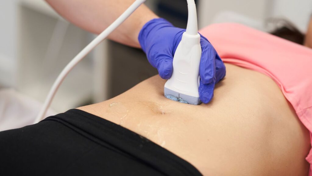
What Is A Pelvic Ultrasound?
A pelvic ultrasound is a safe and easy way to take pictures of the organs in your pelvic area. It is a painless process that uses sound waves. For the test, a small device called an ultrasound probe sends sound waves into your body. These waves bounce off the organs, and the echoes make images on a computer screen. This test helps doctors check the uterus and ovaries’ size, shape, and position.
How It Helps In Diagnosing Female Infertility
By tracking how follicles grow during the menstrual cycle, especially during an IVF cycle, doctors can determine whether ovulation occurs regularly and check how well the body reacts to fertility medications. The size and number of mature follicles provide helpful information about the ovarian reserve and the chance of successfully retrieving eggs. Moreover, ultrasound can help assess hormone levels by linking follicle growth with estrogen levels.
Types of Pelvic Ultrasound Scans
Pelvic ultrasounds can be done in two main ways: through the abdomen (transabdominal) and the vagina (transvaginally). Both methods use sound waves to create images. However, they differ in where the probe is placed and the level of detail they provide. The choice of which method to use depends on several factors. These factors include the specific information needed and how comfortable the patient feels.
Transabdominal Vs. Transvaginal Ultrasound
Transabdominal ultrasound is done by moving an ultrasound probe across the lower abdomen. This gives a general view of the pelvic organs. On the other hand, transvaginal ultrasound involves gently placing a special ultrasound probe into the vagina. This may cause some light pressure but is usually not painful.
When And Why Each Type Is Used
A transabdominal ultrasound is usually the first way to check the pelvic area. It provides a good overview of the organs and can find larger masses or issues. This kind of scan is also helpful for women who are not sexually active or who want a simpler option. However, it does not show as much detail, so it might not be the best choice for tracking follicle development.
In contrast, a transvaginal ultrasound is better for monitoring follicles, which is very important for fertility treatments like IVF and IUI. By watching how the follicles grow, doctors can find the right time for egg retrieval or, for natural conception, the best time for intercourse. Your doctor will tell you which type of ultrasound to have and how often to get it based on your needs and treatment plan. Always feel free to ask your sonographer or doctor any questions you have about the process.
Follicular Scan: Tracking Ovulation Through Ultrasound
A special kind of pelvic ultrasound called a follicular scan is often used in fertility treatments. This scan examines how follicles grow in the ovaries and provides helpful information about when ovulation occurs. Doctors can improve their chances of getting pregnant by knowing how follicles develop.
What Is A Follicular Scan?
A follicular or tracking scan is when doctors use transvaginal ultrasound scans over several days during a woman’s menstrual cycle. This helps them watch how ovarian follicles grow and develop. The scanning usually starts early in the menstrual cycle, around day two or three. The exact timing can change based on personal factors and the advice of the fertility specialist.
How It Helps Monitor Follicle Development And Ovulation
Follicular monitoring is important in fertility treatment. It gives real-time details about how the follicles develop and when ovulation is likely. By watching the changes in follicle size and the ovarian tissue around it, doctors can learn how a woman responds to fertility medications and make changes to improve treatment results. This is especially key during IVF cycles, where careful timing for egg retrieval is vital for success.
Ovulation Tracking with Ultrasound
Ultrasound does more than show the reproductive organs. It is very important for tracking ovulation. This technology gives a clear view of the menstrual cycle. Because of this, both natural conception and assisted reproductive technology can be better timed, which helps to improve the chances of a successful pregnancy.
Role Of Ultrasound In Confirming Ovulation
There are several ways to predict ovulation. One popular method is using ovulation predictor kits that notice the LH surge. However, ultrasound provides clearer proof of ovulation. A set of ultrasound scans can follow the growth and disappearance of the main follicle, showing that the egg has been released. This can be very helpful for women with irregular cycles. It can be hard to find the exact time of ovulation with other methods.
How Ultrasound Helps In Fertility Treatments
The use of ultrasound is very important in fertility treatments. It does more than confirm ovulation; it helps make treatments more successful. For example, in IVF treatment, ultrasound is used for follicular monitoring. This allows doctors to know the best time to retrieve eggs. By watching the growth of follicles, they can give a “trigger shot” to induce ovulation exactly when the eggs are ready to be collected.
Pelvic Ultrasound for PCOS Diagnosis
Pelvic ultrasound is more than just tracking ovulation. It is an important tool for diagnosing Polycystic Ovary Syndrome (PCOS), a hormonal issue that many women have. This test helps doctors see the ovaries clearly and check their structure. With this information, they can find signs of PCOS and tell it apart from other conditions.
Identifying Polycystic Ovaries Through Ultrasound
During an ultrasound, the sonographer checks the ovaries, examining the number and size of follicles. Antral follicle count (AFC) of 12 or more follicles, each measuring less than 10mm, often indicates PCOS.
It’s essential to understand that polycystic ovaries can be found in women without PCOS. Also, not every woman with PCOS will show polycystic ovaries in their ultrasound. That’s why doctors look at various things, such as irregular periods, high androgen levels, and ultrasound results, to make a proper diagnosis of PCOS.
How Ultrasound Helps In Managing PCOS-Related Infertility
Managing PCOS includes making lifestyle changes such as improving diet and exercise. Ultrasound gives doctors useful information about how the condition manifests in the body. Looking at the ovaries and the uterine lining, they can see if lifestyle changes are helping. This information allows them to create treatment plans that fit each person’s needs, which can increase the chance of getting pregnant. It’s important to know that managing PCOS can be complicated. It usually means working with different types of doctors, like gynecologists, endocrinologists, and fertility specialists.
Endometriosis Detection Using Ultrasound
Endometriosis is where tissue like the uterine lining grows outside the uterus. It causes pelvic pain and infertility in women. While pelvic ultrasound isn’t the best test for diagnosing endometriosis, it can help find some specific signs and direct further tests.
How Ultrasound Helps In Diagnosing Endometriosis
Pelvic ultrasound can sometimes find endometriomas. These are cysts that form in the ovaries when endometrial tissue grows. On the ultrasound image, these cysts look like dark, fluid-filled sacs. While endometriomas can suggest endometriosis, not all women with endometriosis have visible cysts on an ultrasound.
Ultrasound can also show other signs of endometriosis, like adhesions. Adhesions are bands of scar tissue that can connect organs. They may appear as fine lines or changes in the normal shape of the pelvic organs on the ultrasound.
Limitations Of Ultrasound In Detecting Mild Endometriosis
The accuracy of ultrasound for diagnosing endometriosis can depend on various things. These include the skill and experience of the sonographer, the type of ultrasound machine used, and the shape of the patient’s body. For example, women with higher body mass index (BMI) might have harder ultrasounds because denser tissue can hide signs of the disease. If an ultrasound doesn’t provide clear results for suspected endometriosis, doctors might suggest other procedures. One option is laparoscopy. This is a small surgery that allows direct viewing of the pelvic organs.
Other Uterine and Ovarian Abnormalities Detected by Ultrasound
Pelvic ultrasound can detect various problems with the uterus and ovaries that may affect fertility. These include issues like PCOS, cysts, and fibroids. It also checks for irregular shapes and other conditions that impact the fallopian tubes.
Fibroids, Cysts, And Structural Abnormalities
Uterine fibroids are non-cancerous lumps that grow in the uterus’s muscle wall. They are often found during pelvic ultrasounds. Most fibroids do not cause problems or affect fertility. However, some can change the shape of the uterine cavity or block the fallopian tubes, making it hard to get pregnant. Ultrasound is useful for determining the size, number, location, and type of fibroids, which helps doctors plan the right treatment.
Ovarian cysts are also common. They are fluid-filled sacs that appear in the ovaries and are usually harmless. Still, certain types, like endometriomas or big cysts that burst, can impact fertility. Doctors use ultrasound to check the cysts’ size, shape, and features. This helps them decide if more tests or treatments are needed.
Their Impact On Female Fertility
The effect of uterine and ovarian problems on fertility can change based on many things. This includes the type, size, and location of the problem. It also depends on the overall health and age of the person. For example, large fibroids that change the shape of the uterine cavity can block embryo implantation. This can raise the risk of miscarriage or infertility. Also, cysts that block the fallopian tubes can stop the egg from meeting the sperm. This hinders fertilization.
In addition to physical blockages, some problems can affect fertility by changing the hormonal balance needed for conception. For instance, some ovarian cysts can disrupt ovulation. This involves regular menstrual cycles and lowers the chances of getting pregnant. It is important to understand that just having an abnormality does not mean that a person will be infertile.
Benefits and Limitations of Ultrasound in Diagnosing Infertility
Pelvic ultrasound is a helpful tool for diagnosing infertility. It is non-invasive, easy to access, and provides live images of the reproductive organs. However, like any medical test, it has its limits. It works best with a full medical history, a physical exam, and other diagnostic tests.
Advantages Of Ultrasound In Fertility Diagnosis
Ultrasound is a major benefit in fertility testing. It lets doctors see the reproductive organs without causing pain or needing surgery. Unlike laparoscopy, ultrasound doesn’t require cuts or anesthesia, making it safer for many women. Plus, it is cheaper than other types of imaging, like MRI, so more people can use it.
Ultrasound provides real-time images, which means doctors can watch important processes, like follicular development and ovulation, happen right before them. This helps during fertility treatments because it allows for the right timing of things like egg retrieval or IUI. Also, ultrasound is very safe and has no known negative effects on fertility, making it a great choice for women trying to get pregnant.
When Additional Tests May Be Required
Ultrasound gives important details about the structure and function of reproductive organs. However, it does not fully show the health of fertility. You often need extra tests, like bloodwork, to check hormone levels. These tests help evaluate ovarian reserve, thyroid function, and other hormonal factors that can affect fertility. For example, high cholesterol, high blood pressure, or diabetes can impact fertility and might need further testing and care.
Also, ultrasound can’t diagnose some problems that may cause infertility, like tubal blockages or pelvic adhesions. A special test called a hysterosalpingogram (HSG) might be needed. This test uses a contrast dye in the uterus and fallopian tubes and X-rays to check if the tubes are open. Even with these limits, ultrasound is a useful first step. It also helps guide further checks and focused treatment plans.
Conclusion
Pelvic ultrasound is crucial for diagnosing female infertility as it provides clear insights into reproductive health. It helps assess factors like ovulation, PCOS, endometriosis, and other conditions that may impact fertility. Understanding the benefits and limitations of ultrasound aids in developing effective treatment plans. Regular check-ups using ultrasound play a key role in evaluating infertility and guiding doctors on the next steps.
For expert fertility care and personalized treatment, visit Ovum Fertility and consult with specialists to take the right steps toward your parenthood journey.
FAQs
1. How often should one undergo a pelvic ultrasound for infertility assessment?
The number of pelvic ultrasounds needed to check infertility depends on the patient. Your GP might suggest having scans every few days during follicular monitoring, especially if you are in an IVF cycle. If you get treatment, the number of ultrasounds can change based on your needs and response. Routine ultrasounds outside of treatment cycles rely on your specific medical history, symptoms, and AFC.
2. Can a pelvic ultrasound detect ovulation problems?
Yes, an ultrasound can monitor follicle development in the ovaries. It helps determine if ovulation is occurring and assesses the health of the follicles, which are necessary for conception.
3. How does a pelvic ultrasound check for uterine abnormalities?
A pelvic ultrasound can detect fibroids, polyps, or abnormalities in the uterine lining. These issues can affect implantation and pregnancy, making the test essential for fertility evaluations.
4. How often is a pelvic ultrasound needed during infertility treatment?
The frequency depends on the specific infertility issue and treatment plan. Some women may need multiple ultrasounds during ovulation tracking or fertility treatments like IVF to monitor egg development and uterine conditions.